
Discoveries
The Religious Brain Project
Billions of people make important life decisions, frame core beliefs, and participate in social communities based on spiritual and religious experiences. How the brain experiences these feelings is a critical, understudied process with implications for how religious convictions are developed and maintained. The results of the first study of the Religious Brain Project are now published in Social Neuroscience
Press:
Top of Mind with Julie Rose: BYU Radio
Robert Kirby: The Salt Lake Tribune
A Brain Mechanism for Pediatric Bipolar Disorder
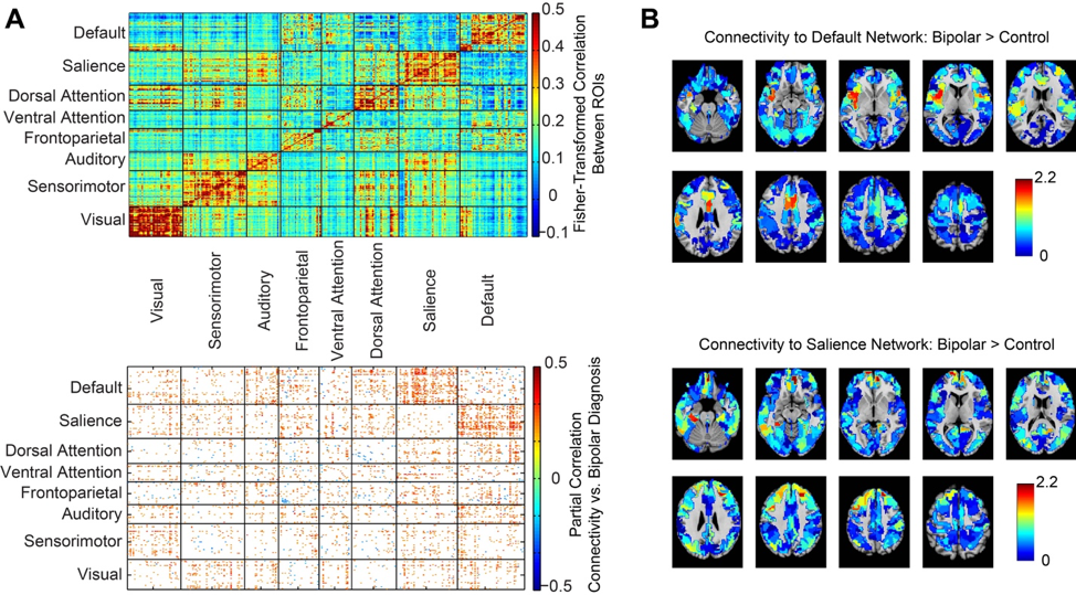
Although pediatric bipolar disorder is among the most disabling psychiatric conditions affecting youths, little is known about how brain connections may be altered. In studying brain images of 80 youths (32 with bipolar disorder, 48 healthy control), we found participants with bipolar disorder showed abnormal development during adolescence of negative or inhibitory connections between two important brain networks that regulate attention to the outside world vs. one’s internal thoughts and narrative. We find that such negative connections comprise the most abnormal brain connections in pediatric bipolar disorder, and that delayed or abnormal development of such negative connections may be a core feature of how bipolar disorder forms in the brain. These abnormal connections are concentrated between 2 brain networks: the default mode and salience network. You can read the full story here:
Abnormal functional connectivity between default and salience networks in pediatric bipolar disorder
Short Sleepers?
Sleep is one of the biggest mysteries in science. Nobody really knows for sure why we sleep. But it's an enormous risk to spend a third of our lives immobilized, and almost all animals do it. Interestingly, up to a third of people get quite a bit less sleep than the rest of us, less than 6 hours, and some people only 2 or 3 hours a night. Many of these people feel that they are not tired, and that's all the sleep they need.
The leading theory as to why we sleep is that it is a period of time where short-term memories can be moved to long-term storage in the brain. We replay the day's events in our minds and make copies of these events in areas of the brain that can form more permanent memories. This is called memory consolidation.
Our study is the first that gives evidence for an exciting hypothesis about why some people may need less sleep. We analyzed data from the Human Connectome Project that scanned people while they were supposedly awake, but resting. People who reported less than 6 hours a sleep per night on average showed brain activity patterns that looked more like sleep than those who didn't. So either these people (especially ones who don't feel tired during the day) are able to take little micronaps when stimulation is withdrawn, or they are able to perform tasks like memory consolidation while they are still awake, reducing the need for sleep.
This superpower may come with a downside too. If they are really falling asleep whenever they are not stimulated, they may be more distracted during tasks like driving, and may be at risk that they don't know they have. They may not be aware they are tired, but yet are taking small brief naps or something like it during the day.

Wired for Gaming
We collaborated with Doughyun Han, a psychiatrist from South Korea, to look at what brain connectivity changes we could see in compulsive video game players. Doug has been collecting imaging cases for years, and we were able to analyze over 150 adolescents in the largest sample in the literature of video game players.
The results are now published in 2 studies. One study looks at resting state functional connectivity and the other study examines gamers while playing a simple card game. We find that gamers have stronger connectivity between auditory and motor cortex to attentional regions like the frontal eye fields and salience network. To us these look like training effects, and may be adaptive. We also see over-connectivity between the default mode and salience networks, which is more ominous, as this can be associated with poor executive function, distractibility, and poor concentration.

Common Technique for fMRI is an Artifact?

Brain imagers have looked to other fields for exciting new methods for finding meaning in brain images. One example is the use of tools for looking at time series from economics. For decades, mathematicians have used sophisticated models to learn about how past events in the stock market may predict future performance. A mathematical model called Granger Causality has been ported to predict which brain regions might influence other brain regions. In its most common form, scientists target a few small brain regions to use this technique. We looked at large public databases with over a thousand participants' brain fMRI scans and found that using this technique, we recovered a map of blood flow in the brain, with "sources" near brain arteries and "sinks" near brain veins. A little disappointing, but it's always good to know when your methods may be measuring blood flow when you think they are measuring something more profound, like brain activity. And identifying a problem may be the first step to finding a way around it. The research can be found in PLoS One.
Discussion:
http://neuroconscience.com/tag/granger-causality/
http://www.russpoldrack.org/2013/12/a-discussion-of-causal-inference-on.html
Left- and Right- Dominant Brain Networks

The popular conception that some people are left-brain dominant and some are right-brain dominant is everywhere in popular culture, but has never been accepted by the neuroscience community. We tested left-dominant and right-dominant brain networks in over 1000 individuals using functional connectivity MRI and found that individuals do not exhibit hemispheric dominance, but rather individual subnetworks operate independently. So some individuals may exhibit strong left-dominance in some connections or local networks, but not in other networks. Findings are published in PLoS One.
Press:
http://healthsciences.utah.edu/blog/postings/august_2013/082013_leftbrain.php
http://www.prevention.com/mind-body/emotional-health/left-and-right-brain-personalities-debunked
http://www.huffingtonpost.com/2013/08/19/right-brain-left-brain-debunked_n_3762322.html
http://www.foxnews.com/health/2013/08/19/left-brained-right-brained-personalities-are-not-real-study-shows
http://now.msn.com/left-brain-right-brain-theory-debunked-by-university-of-utah-neuroscientists
http://www.wnyc.org/shows/bl/2013/aug/20
http://gawker.com/the-left-brain-right-brain-distinction-is-as-fake-as-it-1153790191
http://www.youtube.com/watch?v=yE6VTvxkhFs&list=TLaQ1_uYO-c-w
Autism

We have used functional connectivity MRI as a marker for autism and found that in individuals under 20 years of age, an 8-minute MRI scan can be 80-90% accurate as a predictor of whether someone has autism or not. By looking inside the "wiring diagram" of autism, we are learning which networks function abnormally and how we may be able to predict prognosis or monitor treatment in individuals with autism. These results are online at the journal Brain. Early efforts to expand this research to a broad, multisite dataset are found in the journal Frontiers in Human Neuroscience.

The left and right hemispheres of the brain are abnormally connected in autism. The connections that are most abnormal are those that involve brain regions known to process functions that are abnormal in autism, such as recognizing faces, social interactions, and attention. Results are now online at Cerebral Cortex:
Press:
https://sfari.org/news-and-opinion/news/2013/study-seeks-autism-biomarkers-in-brain-imaging-database
http://www.ksl.com/?nid=148&sid=12798200
http://www.sciencedaily.com/releases/2010/10/101013082818.htm
http://www.deseretnews.com/article/700073202/MRI-may-be-diagnostic-tool-for-autism.html
http://news.health.com/2010/10/14/mri-might-screen-for-autism/
http://www.physorg.com/news/2010-10-autism-mri-closer.html
Attention
At any moment there are many things one can pay attention to such as sights, sounds, and internal thoughts. We show that in areas of the brain that process attention, there is a map of the world, with subregions for each of the senses. This allows a “wiring diagram” for control of attention that can explain how we can switch our attention from one thing to another. Read the details now online at Proceedings of the National Academy of Sciences:
Press:
http://www.sciencedaily.com/releases/2010/11/101101151724.htmhttp://esciencenews.com/articles/2010/11/01/utah.researchers.discover.how.brain.wired.attention

Map of “attentional space” in the brain.